On a January afternoon in 1953, Maurice Wilkins walked James Watson into a room at King's College London and showed him a photograph. It was an X-ray diffraction image — technically labelled number 51 — of a DNA fiber at near-maximum humidity, taken the previous May. Watson later wrote that his jaw fell open the moment he saw it.1 The dark X-shaped pattern at its center told him everything he needed: the molecule was a double helix. Rosalind Franklin, who had spent 100 hours coaxing that image out of a strand of DNA finer than a human hair, was not in the room. She did not know the photograph had been shared.
Early Life
Franklin arrived at the Laboratoire Central des Services Chimiques de l'État in Paris on 14 February 1947, one of fifteen researchers in Jacques Mering's group.2 She was 26, freshly credentialed from Cambridge with a doctorate in physical chemistry earned partly through wartime coal research, and she spoke fluent French. Mering was one of the few scientists applying X-ray diffraction not to perfect crystals but to disordered, amorphous substances — exactly the kind of messy matter that biological molecules would later prove to be. He taught Franklin to interpret what others saw as noise. Biographer Anne Sayre later reported that the Paris years were the happiest period of Franklin's life3: she hiked on weekends with colleagues, argued passionately about science over coffee, and published five landmark papers on graphitizing and non-graphitizing carbons — a distinction still standard in materials science today3. When she finally left France in 1950, reluctantly, she carried the technique that would make her indispensable to the DNA problem.
Career
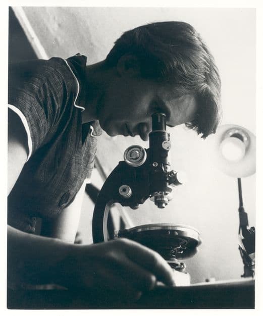
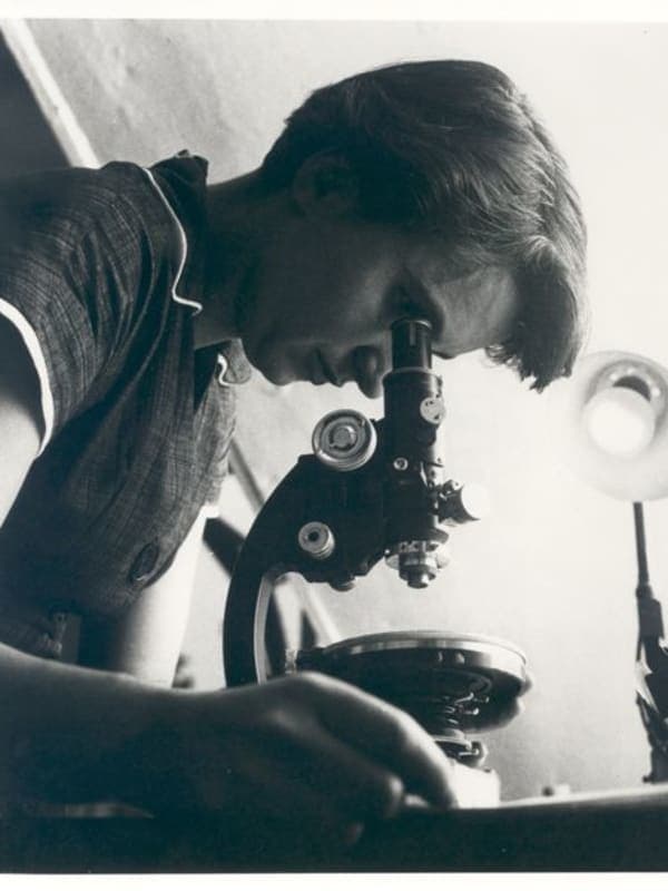
In May 1952, inside the basement of King's College London, Franklin and her graduate student Raymond Gosling suspended a DNA fiber inside a camera she had designed herself and fired an X-ray beam at it for one hundred hours.4 The humidity in the chamber was held at roughly 92 percent5 — the precise condition, Franklin had determined, that coaxed DNA into its wet B form. The resulting image, the fifty-first diffraction photograph Gosling had taken6, showed a crisp black X on a pale background. It encoded the helix's dimensions, pitch, and the offset between its two antiparallel strands. Franklin set it aside: she was focused on the drier A form, which she believed would yield a more direct mathematical solution. By January 1953 she had drafted manuscripts concluding that both forms carried a double-helical backbone. Her A-DNA papers reached Copenhagen on 6 March 1953 — one day before Watson and Crick completed their model at Cambridge.2
What Franklin did not know, as she prepared to leave King's for Birkbeck College, was that Wilkins had shown Watson Photo 51 without her knowledge or permission.6 Watson recognized the pattern immediately, because Crick had previously described what a helix's diffraction signature would look like. Watson and Crick published their double-helix model on 25 April 1953.7 In the same issue of Nature, Franklin and Gosling's paper appeared third — positioned, through editorial arrangement, to look like supporting evidence for a discovery it had actually preceded and enabled. Watson and Crick's paper acknowledged only a "general knowledge" of Franklin's unpublished work.1 Franklin's data corroborated the model; it is not clear she knew how thoroughly it had built it.
At Birkbeck, freed from the hostility of King's and under the directorship of crystallographer J. D. Bernal, Franklin pivoted to viruses. Working with future Nobel laureate Aaron Klug, she applied the same rigor she had brought to DNA — designing new sample-preparation methods, building custom equipment, spending months on the quality of a single image before publishing. In 1955 she published the first three-dimensional structural model of any virus, derived from X-ray analysis of the tobacco mosaic virus.7 A year later she reported that the virus's RNA was embedded in a spiral within a hollow tube of protein.7 In the fall of 1956, she was diagnosed with ovarian cancer.1 She continued working through three operations and experimental chemotherapy1, on tobacco mosaic virus and then on polio. Her team concluded that the polio capsid has icosahedral symmetry — sixty-sided, like a soccer ball.8 She worked until several weeks before her death on 16 April 1958, at thirty-seven.1
Legacy
Watson, Crick, and Wilkins shared the 1962 Nobel Prize in Physiology or Medicine.9 Franklin had been dead for four years; the Nobel is not awarded posthumously. None of the three gave her credit in their acceptance remarks.9 Franklin's role might have remained a footnote had Watson not caricatured her a decade later in The Double Helix, presenting her as a bad-tempered bluestocking incapable of interpreting her own data9. The book was widely read and widely condemned, and it dragged Franklin's contributions into the open. In 1975, her friend Anne Sayre published an angry rebuttal biography; by the 1980s, historians of science were reconstructing exactly what Photo 51 had meant to the race for the double helix. Francis Crick later acknowledged, "I'm afraid we always used to adopt — let's say, a patronizing attitude towards her."2 James Watson eventually conceded that, had she lived, Franklin would ideally have been awarded a Nobel Prize in Chemistry.2 In 2004, a medical university in North Chicago renamed itself the Rosalind Franklin University of Medicine and Science, taking Photo 51 as its logo.2 In 2019, the European Space Agency named its ExoMars rover Rosalind Franklin8 — a machine built to drill into the Martian surface in search of the molecular signatures of life.